
Fig. 7.
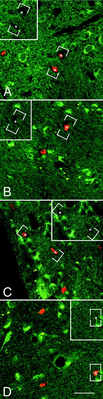
The newly generated cells in the parenchyma of the adult rat forebrain do not express TrkB receptor after coinfusion of BDNF and BrdU. A–D, Representative fluorescent photomicrographs of coronal sections, captured by confocal microscopy, showing the distribution of the nuclei of newly generated BrdU+ cells and the cells expressing the full-length TrkB receptor in the striatum (A), septum (B), thalamus (C), and hypothalamus (D). The BrdU+cells were identified with a rhodamine-conjugated secondary antibody (bright orange), and the TrkB was visualized with a fluorescein-conjugated secondary antibody (green). The sections were visualized with either a dual fluorescein–rhodamine filter or with only a fluorescein filter (insets). In none of the four regions analyzed did the BrdU+ cells (e.g., asterisks) express the TrkB receptor. Frequently, however, the BrdU+cells were adjacent to TrkB+ cells. The absence of double-labeled cells suggests that the BDNF may have an indirect effect on the proliferation and/or survival of newly generated cells. Scale bar: A–D, 30 μm.