
Fig. 2.
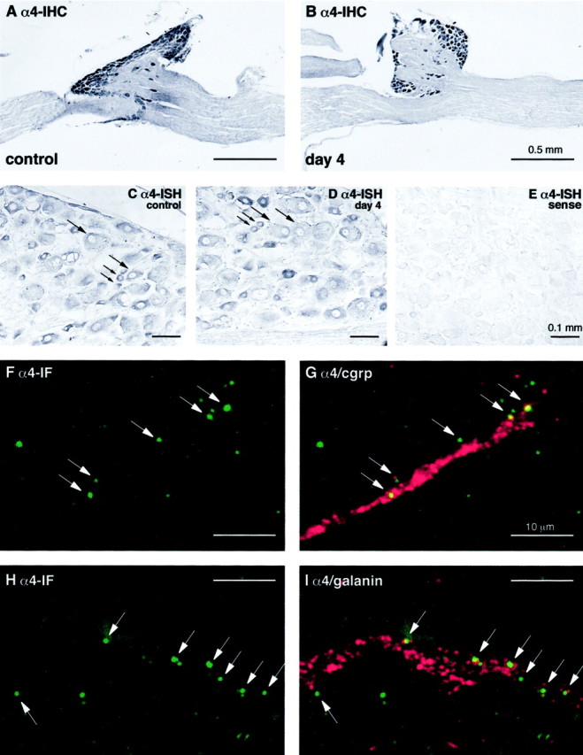
α4 integrin expression after sciatic nerve injury in mouse and rat. A and B show α4 immunoreactivity visualized by immunoperoxidase in the L5 spinal ganglia 4 d after axotomy of the mouse sciatic nerve. Note that α4 is expressed within the DRG neurons on both the control, unlesioned (A), and lesioned (B) side, without any changes in expression level. C–E show the effects of axotomy on α4 mRNA levels in rat sciatic nerve, as assessed by in situhybridization on sections of DRG neurons from the experimental (C, E) and control (D) side.C and D show the expression of α4 mRNA in both large- and small-diameter neurons (large andsmall arrows, respectively) both before (C) and 4 d after axotomy (D). E shows a sense control with no nonspecific hybridization. F–I represent higher power confocal images showing α4 immunoreactivity (green) and either CGRP or Galanin (red) in the distal stump 4 d after sciatic nerve crush. These show the presence of discrete foci of α4 associated with growth cones marked by CGRP or galanin immunoreactivity, confirming the presence of this integrin in regenerating nerves. Note that some of the α4 immunoreactivity in these confocal images is seen at the edge of the CGRP–galanin-labeled areas, as would be expected given the cell surface expression of α4 integrin and the cytoplasmic localization of CGRP or galanin within the growth cones.