
Fig. 3.
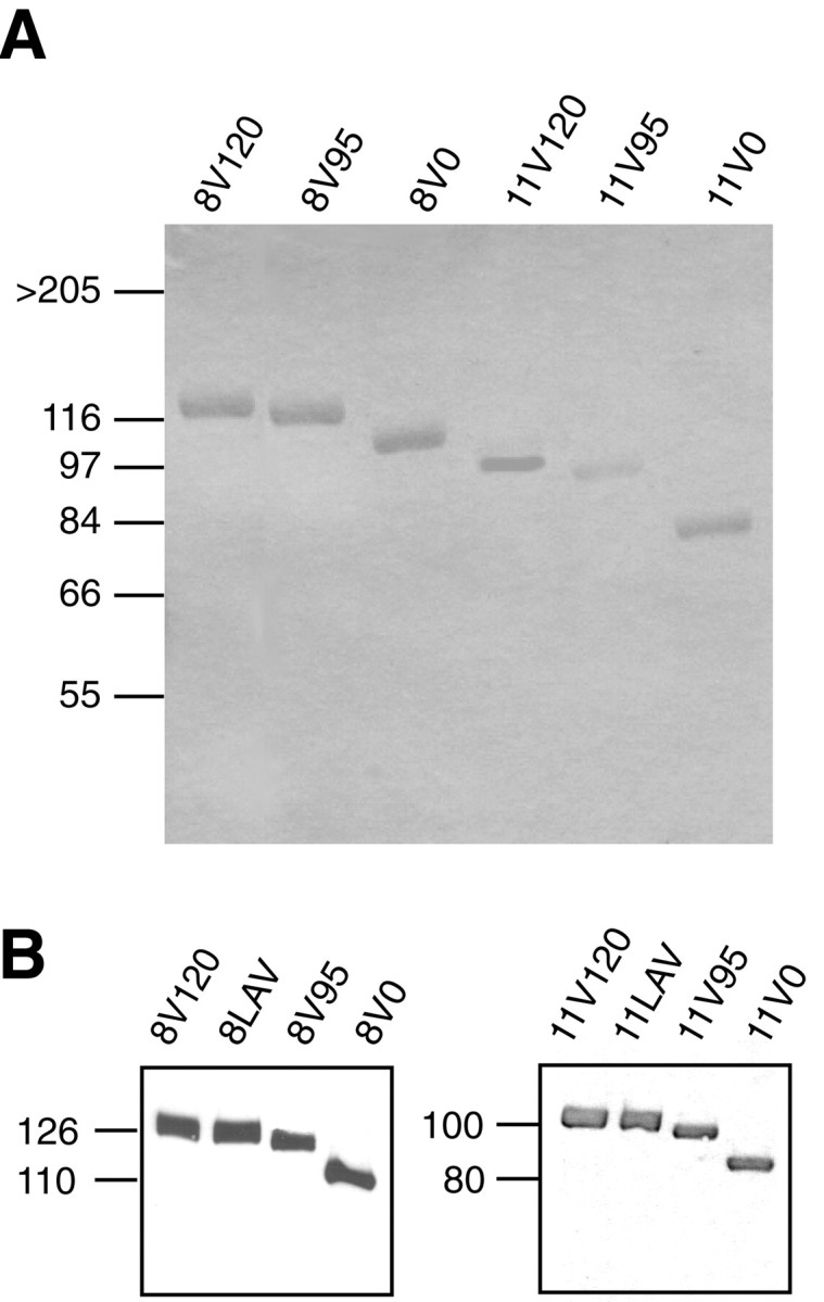
SDS-PAGE and immunoblot analyses of recombinant fragments. A, Recombinant FN fragments purified on protein A-Sepharose columns, as described in Materials and Methods, were subjected to SDS-PAGE under reducing condition and visualized by Coomassie staining. Molecular weight markers are shown on theleft. Note that all fragments appear as single bands.B, Purified FN fragments (0.5 μg/lane) were subjected to SDS-PAGE under reducing conditions followed by immunoblotting with polyclonal anti-Fc antibody. Left, 8–15 fragments.Right, 11–15 fragments.