Fig. 5.
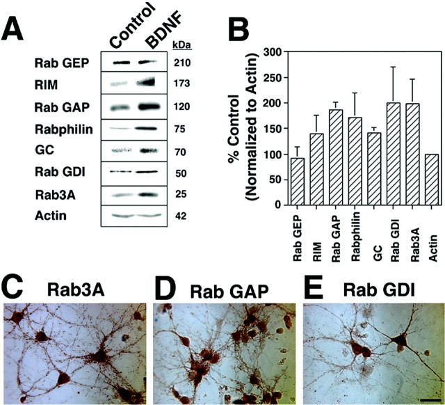
BDNF induces translation of identified genes and their regulatory proteins in hippocampal neurons. A, Equal amounts (50 μg) of protein from control and BDNF-treated (6 hr; 50 ng/ml) cultures were loaded in each lane, electrophoresed, immunoblotted with antibodies, and visualized with ECL. GC (70 kDa) and Rab3A (25 kDa) are upregulated by BDNF treatment. Several regulatory proteins were also induced by BDNF: RIM (173 kDa), Rab GAP (120 kDa), Rabphilin (75 kDa), and Rab GDI (50 kDa). Rab GEP (210 kDa) was unaltered. Actin (42 kDa) levels remained unchanged. B, Quantitation of increase in protein levels in BDNF-treated samples relative to control cells after normalization to actin is shown (n = 4 except for GC, n = 3, and Rab GEP, n = 2). C–E, Identified proteins are localized to neurons. Hippocampal cultures were fixed and immunostained using avidin–biotin–horseradish peroxidase visualization. Rab3A (C) appears in a punctate pattern in neuronal processes. Rab GAP (D) and Rab GDI (E) staining is also localized to neurons. Note the cellular heterogeneity of staining intensity and localization to pyramidal-like neurons. Scale bar, 50 μm.

