
Fig. 7.
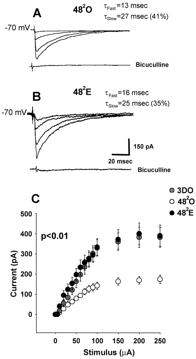
In estrogen-treated animals at the 482 time point, synaptically evoked IPSC amplitude has recovered to baseline values and is greater than that in oil-treated controls. Recordings were made with a CsCl internal solution. A, Representative individualtraces of IPSCs evoked in a 482O cell using 50, 100, 150, and 250 μA stimulating currents.B, Representative individual traces of IPSCs evoked in a 482E cell using the same stimulus intensities as in A. Evoked currents are blocked by bicuculline. τdecay fast and τdecay slowvalues in A and B apply specifically to the cells shown. C, Averaged stimulus–response curves for 3DO (gray circles; n = 12 cells from 6 animals), 482O (white circles; n = 20 cells from 10 animals), and 482E (black circles;n = 22 cells from 10 animals) groups. There is no difference in peak IPSC amplitude between 482E and 3DO groups (p > 0.1), whereas IPSC amplitude is significantly reduced in the 482O group compared with both 482E and 3DO groups (p < 0.01).