
Fig. 9.
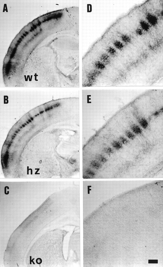
5-HT immunocytochemistry in brains of VMAT2 wt (A, D), hz (B,E), and ko (C, F) mice assessed at P7. A–C, 5-HT immunostaining of coronal sections reveals the barrel pattern in wt and hz mice; no 5-HT immunostaining is visible in VMAT2 ko mice (C). D–F, Higher magnification of sections shown in A–C. Scale bar (shown in F): A,B, 400 μm; C, 430 μm;D–F, 130 μm.