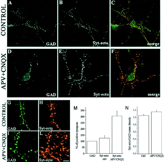
Fig. 4.
Blockade of glutamate receptors affect glutamatergic, but not GABAergic, presynaptic nerve terminals.A–F, Immunofluorescence images of control (A–C) and APV–CNQX-treated (D–F) 15-d-old hippocampal neurons incubated for 5 min in the presence of Syt-ecto Abs (B,E) and counterstained with antibodies against the synthetic enzyme GAD (A, D). In control neurons, the few synapses labeled by Syt-ecto Abs are generally GAD positive (A, B, see also merged image inC). In APV–CNQX-treated neurons, the synapses positive for Syt-ecto Abs (E) largely exceed GABAergic terminals (D) (see merged image inF). G–L, Details of control (G, H) and APV–CNQX-treated (I, L) neurons stained for Syt-ecto Abs (H, L) and for GAD (G,I). Scale bar: A–F, 30.7 μm;G–L, 10.2 μm. M, Histogram showing the quantitative analysis of the percentages of Syt-ecto Ab-positive synapses in control and APV–CNQX-treated neurons, normalized to the number of GAD-positive synapses. N, Histogram showing that the amount of internalized Syt-ecto Abs is not significantly different in GABAergic terminals of control and treated neurons. Syt-ecto Ab intensity values are normalized to GAD immunoreactivity.

