
Fig. 6.
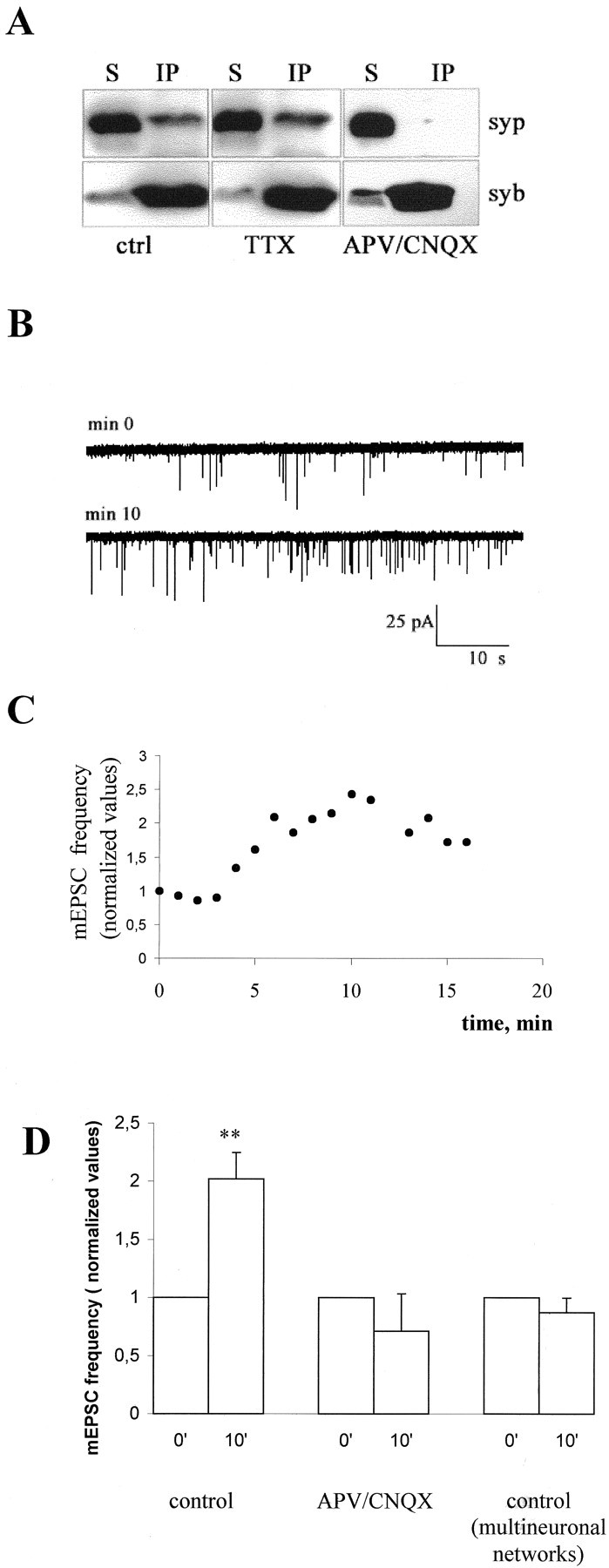
Impairment in the formation of the synaptophysin-synaptobrevin–VAMP2 complex is detectable in APV–CNQX-treated neurons and is responsible for the increase in mEPSC frequency. A, Triton X-100 extracts of control and APV–CNQX-treated 12-d-old neurons were immunoprecipitated using the monoclonal antibody against synaptobrevin–VAMP2 (syb). Immunoprecipitates (IP) and their corresponding supernatants (S) were analyzed using polyclonal antibodies against synaptophysin (syp). Note that synaptophysin is efficiently immunoprecipitated from control cultures and is almost completely detectable in the supernatant of APV–CNQX- treated cultures. B, Representative recordings from a single 12-d-old hippocampal neuron forming autaptic contacts, intracellularly perfused via the patch pipette with a peptide corresponding to the 32-residue-long N-terminal segment of synaptobrevin–VAMP2, which inhibits complex formation. C, Time course of the increase in the frequency of mEPSCs recorded from the same neuron as inB. D, Histogram showing the increase in mEPSC frequency occurring in control neurons 10–11 min after the beginning of recordings (p < 0.002). No effect is detectable in either single neurons maintained in APV–CNQX (p > 0.1) or in neurons grown in polyneural networks (p > 0.1). Values are normalized to the first or second minute of recording.