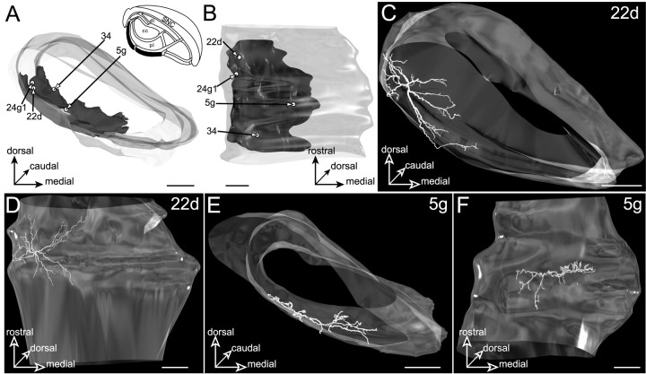
Fig. 15.
Neurons located in the ventral lamina (filled area, inset inA) innervated by the striatal sector related to visual cortical areas. The SNR is shown from a rostral view inA and a ventral view in B.C–F, The dendritic arborizations of neurons22d and 5g. The neurons are examined from a rostral view in C and E and a ventral view in D and F. Note that the neurons display flat dendritic fields spreading along the ventral border of the SNR. Scale bars, 350 μm.

