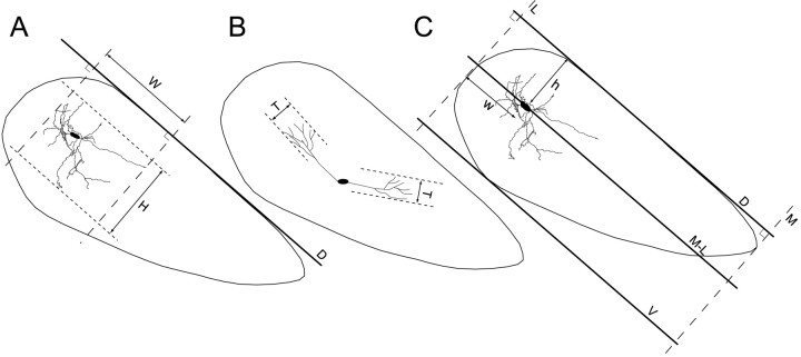
Fig. 2.
Coronal sections of the left SNR illustrating the axis of reference used to measure the length of the dendritic arborizations and to determine the coordinates of the somata of the labeled neurons within the SNR. A, The mediolateral (W) and dorsoventral (H) extent of the dendritic fields were measured, relative to the D axis passing along the dorsal edge of the SNR. This axis takes into account the inclination of the SNR into the brain and allows the definition of the largest mediolateral axis of the nucleus. B, Illustration of the method used to measure the thickness (T) of neurons presenting a discoid dendritic field (see Materials and Methods for details). C, The dorsoventral and mediolateral coordinates of the neuronal somata within the SNR were determined using the axes D, M-L, V,M, and L. These axis were traced on the coronal section of the SNR that contains the soma of the neuron studied. The axis D passes along the dorsal surface of the SNR and defines the mediolateral axis of the nucleus. The axisM-L parallels the axis D and passes through the soma of the neuron. The axis V parallels the axis D and passes tangentially to the ventral surface of the SNR. The axis L is orthogonal to the axisD and passes tangentially to the lateral edge of the SNR. Finally, the axis M is orthogonal to the axisD and passes tangentially to the medial edge of the SNR. The dorsoventral coordinate of the neuron (h) was determined by measuring the distance between the axisM-L and the axis D. To compare the position of neurons from different animals, this value was normalized relative to the maximal thickness of the SNR measured as the distance between the axis D and the axis V. The mediolateral coordinate (w) was determined by measuring the distance between the soma of the neuron and the axisL. This measure was normalized relative to the maximal extension of the nucleus along its mediolateral axis. This extension was determined as the distance separating axes L andM.

