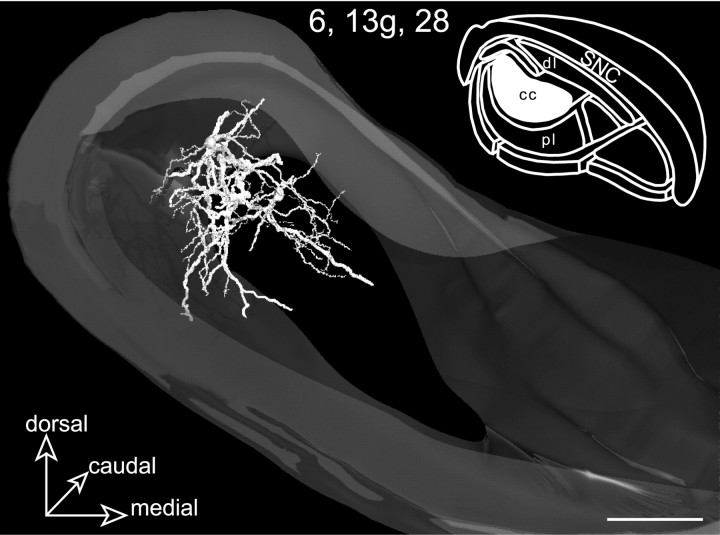
Fig. 6.
Rostral view of a 3D reconstructed model of the SNR incorporating the neurons 6, 13g, and28 labeled in the central core (cc). Remarkably, the overall dendritic arborizations formed a core structure that perfectly matched the projection field of the orofacial striatal sector (inset, filled area). Scale bars, 350 μm.

