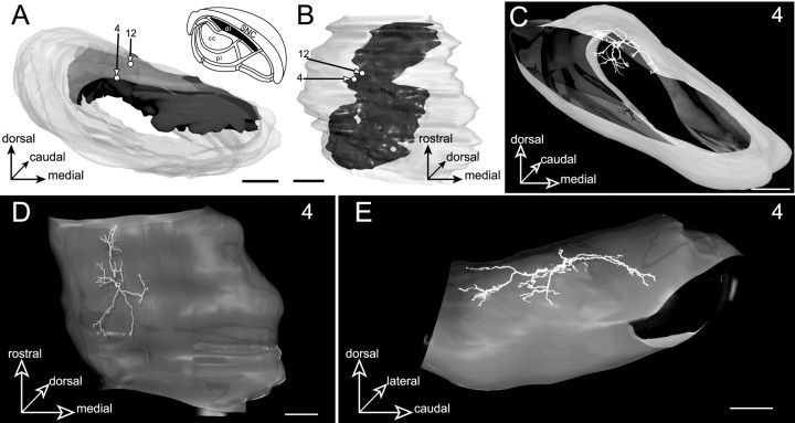
Fig. 7.
Neurons located in the dorsal lamina (dl, filled area, inset inA) innervated by the striatal sector related to insular, gustatory, and perirhinal cortical areas. A,B, Three-dimensional reconstruction of the SNR illustrating the position occupied by the somata of the labeled neurons (4 and 12) with respect to the striatal projection field. The SNR is examined from a rostral view inA and from a ventral view in B.C–E, The 3D reconstructed dendritic arborization of neuron 4. The labeled neuron is examined from a rostral view inC, a ventral view in D, and a medial view in E. Note the similitude between the geometry of the dendritic arborization and the spatial organization of the striatal projections. Scale bars, 350 μm.

