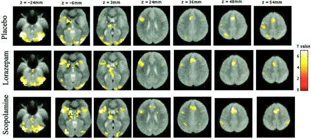
Fig. 3.
Regions showing activations during word-stem completion (new and old word-stems) against fixation, as identified by random effects analysis in the placebo, lorazepam, and scopolamine groups, respectively. Activations are rendered on transverse mean normalized EPI images (EPI images averaged over several volunteers; threshold = p < 0.001). EPI images are used for anatomical descriptions, because they show the same distortions as activation data. A pathway of regions is activated, including bilateral extrastriate regions, left frontal regions along the inferior frontal gyrus, the anterior cingulate, and bilateral cerebellar cortex. The left lateral parietal cortex and precentral gyrus were only significantly activated in the placebo group.

