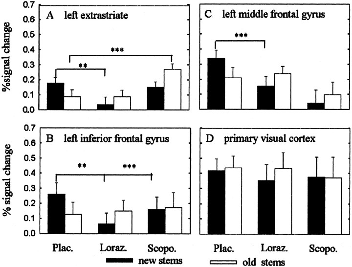
Fig. 5.
Plots of percentage of signal change. Mean and SEM as a function of group and repetition (new stems, black symbols; old stems, white symbols). The plots inA–C derive from the maximum voxel in each region identified by random effects analysis. D shows a voxel in primary visual cortex. The placebo group shows repetition suppression in left extrastriate, left inferior frontal, and left middle frontal cortices. There is an absence of repetition-related reductions in all three brain areas after lorazepam and scopolamine. Compared with placebo, lorazepam reduced activations to new words in all three brain regions. In contrast, scopolamine showed increased activations to old words compared with placebo in extrastriate cortex. Note that the voxel in primary visual cortex shows neither repetition suppression nor an effect of drug. **p < 0.05; ***p < 0.001; ANOVAs followed by post hoc Tukey's tests comparing activations to new and old words between placebo and drug groups.

