Fig. 6.
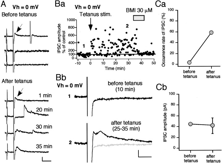
LTP of disynaptic IPSC is mediated by an increase in the occurrence rate of GABA release. A, Sample traces before (top 3 traces) and after tetanus (bottom 4 traces). Membrane conductance was monitored with a test pulse (2 mV, 3 msec) throughout the experiment. IPSCs were obtained at a holding membrane potential of 0 mV, at which EPSCs were nullified. Before tetanus, an IPSC indicated by a small arrow was recorded only once. After tetanus, multiple IPSCs were evoked in almost all sweeps, including the same IPSC as the one seen before tetanus. Calibration: 50 pA, 50 msec. Ba, The afferent fibers were tetanized at time 0 (arrow). Peak ensemble IPSC amplitudes measured within 50 msec after test stimulus are plotted against time for a single cell. BMI, applied during the time indicated by the gray bar, abolished all IPSCs. Averaged sweeps recorded at the times indicated (1, 2) are shown in Bb. Calibration: 10 pA, 20 msec.Ca, Comparison of the occurrence rate of the IPSC indicated by an arrow in A between before and after tetanus. The occurrence rate dramatically increased after tetanus. Cb, Amplitude of the IPSC remained almost the same.

