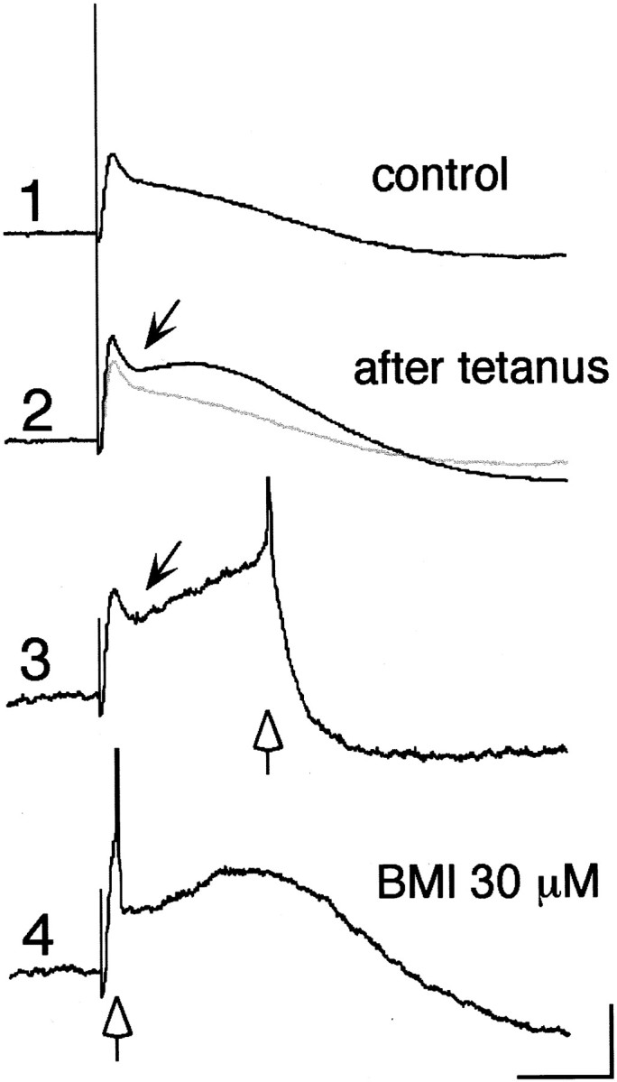
Fig. 8.

Simultaneous LTP of EPSP and disynaptic IPSP. Evoked synaptic potentials before (1) and 45 min after (2, 3) tetanic stimulation (100 Hz, 1 sec, 500 μA, 250 μsec) and after BMI application (4). Note that EPSP as well as a hyperpolarizing component (arrow) was significantly potentiated (2) and that a spike was generated occasionally after hyperpolarizing deflection (3). BMI abolished the hyperpolarizing component, so that a spike was triggered synchronously at the timing of a test stimulus (4). Open arrows in3 and 4 indicate the timing of spike generation. Calibration: 5 mV, 50 msec.
