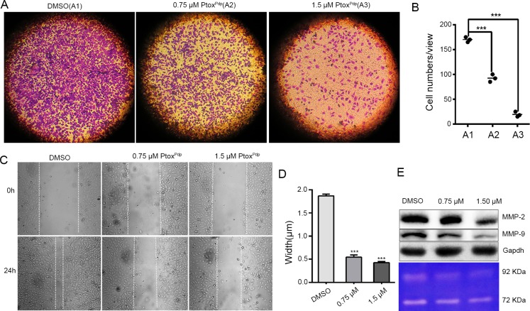
Fig 4. Inhibition of liver cancer cell migration and invasion by PtoxPdp.
(A) Inhibition of HCCLM3 cells migration by PtoxPdp. (B) Inhibition of HCCLM3 cell invasion by PtoxPdp and quantification. The invasive cells were stained with crystal violet. The results were expressed as invasive cell numbers per field of view (mean± 5 SD, n = 6). ***P < 0.001 compared with DMSO-treated group. (C) Wounded HCCLM3 cells were treated with 0, 0.75, or 1.50 μM PtoxPdp for 18 h. (D) Quantitative statistics analysis of the width of gaps in the wound assay in (C). ***P < 0.001 compared with DMSO-treated group. (E) Western blotting (top) and gelatin zymography (bottom) analyses of matrix metalloprotease inhibition, under the indicated conditions. The object size: 10×10 in Fig 4A and 20×10 in Fig 4C.

