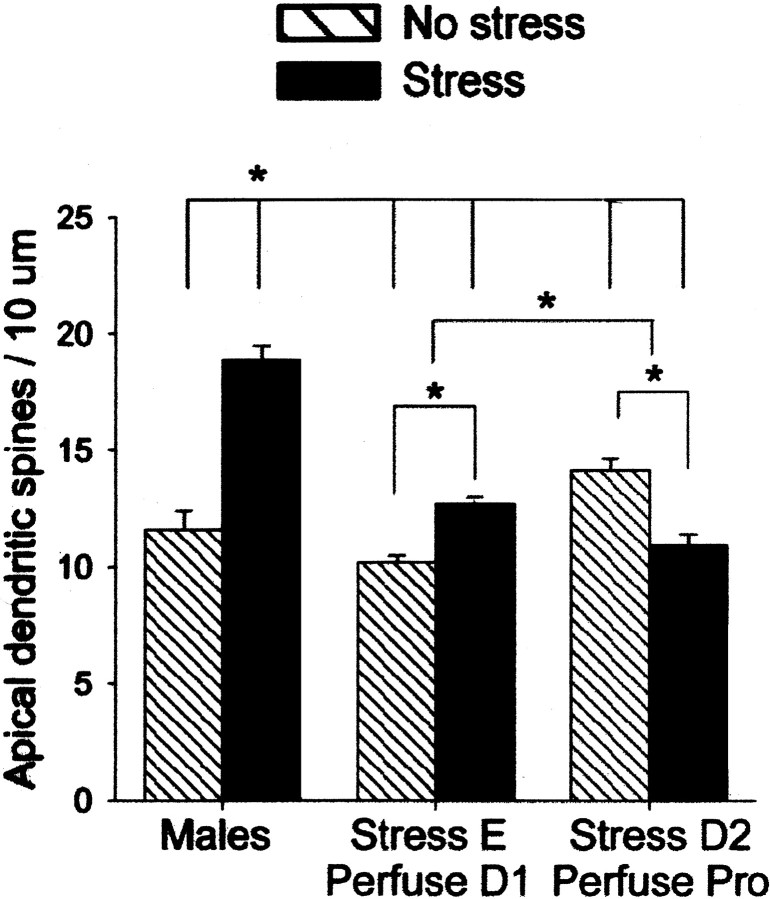
Fig. 2.
Opposite effects of stress on spine density in males versus females. Graph illustrates the mean (±SEM) density of apical dendritic spines on pyramidal cells in area CA1 of the hippocampus 24 hr after exposure to an acute stressor of brief inescapable tail stimulation. Significant differences are noted withasterisks. Under unstressed conditions, females in proestrus had a greater density of spines than males or females in diestrus 1. Exposure to the stressor increased synaptic spine density in males and decreased density in females that were stressed in diestrus 2 and perfused 24 hr later in proestrus.

