Fig. 6.
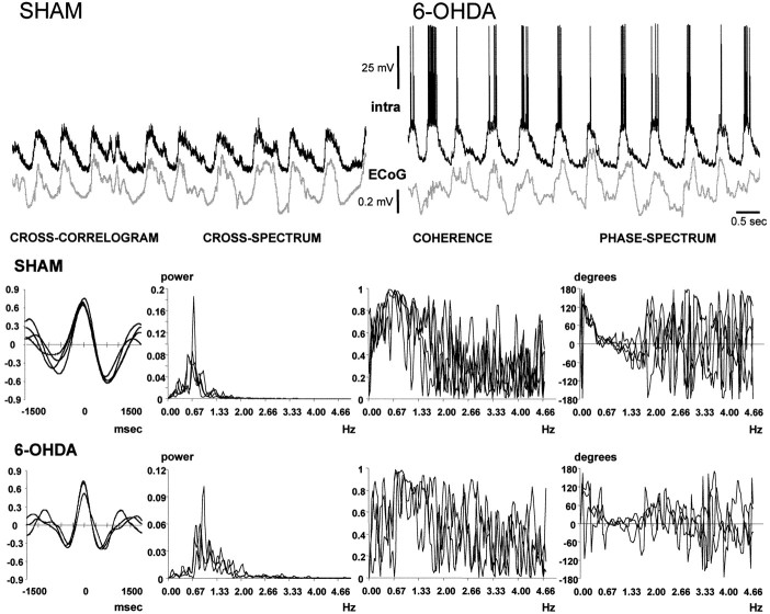
Visual inspection of simultaneous recordings of the frontal electrocorticogram and the membrane potential of striatal neurons indicated that the two waveforms were oscillating synchronously at ∼1 Hz. The signals are displayed as they were recorded, but note that they were down-sampled, smoothed, and standardized before analysis. The belief that the signals were synchronized was substantiated through the analysis of cross-correlograms and by means of coherence analysis. Each row of graphics shows the cross-correlogram, cross-spectrum, coherence spectrum, and phase-spectrum corresponding to the signal pairs displayed in the upper part of the figure (top row of graphics, sham-lesioned rat;bottom row of graphics, rat with nigrostriatal lesion). In each graphic, the results of the analysis of several 30 sec epochs from the same signal pair were superimposed. The four disjoined 30 sec epochs depicted for the sham-lesioned rat were chosen from a recording session elapsing 15 min. A 12 min recording session from a 6-OHDA-lesioned rat provided the three 30 sec epochs that were chosen for the bottom row of graphics. Both signal pairs showed strong correlations. The cross-spectra revealed a powerful common frequency component of ∼0.7 Hz (SHAM) or ∼0.9 Hz (6-OHDA), and the signals displayed a very high coherence and a lineal phase relationship at the dominant frequency.

