Fig. 1.
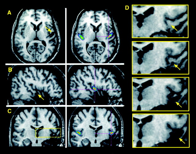
MRI scan of a patient (RTA3 in Table 1) with a removal in the right HG in whom the excision includes the anterolateral 50–60% and the undercutting extends to 60–70%. The MRI scan is transformed into standardized stereotaxic space; illustrated are planes of section oriented horizontally (A; z = 4), sagittally (B; x = 46), and coronally (C; y = −17). The left panels of A–C show the patient's scan alone, with an arrow indicating the region of excision–undercutting. The right panels ofA–C show the patient's scan coregistered with an anatomical probabilistic map of HG derived from normal individuals (Penhune et al., 1996); the map is scaled to show voxels that have a 25% or greater probability of lying within HG. Thecrosshairs indicate the same position in standardized space as the arrow. Note the correspondence between the position of HG as determined from the map and the patient's partially excised HG region. The yellow box in Cindicates the region of the removal pictured in close-up inD, which illustrates the transition from intact, to undercut, to fully excised tissue (coronal sections taken at 3 mm intervals; posterior to anterior; from y = −23 to −14). Arrows again correspond to thecrosshairs in the other panels and indicate the location of HG region.

