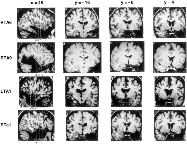
Fig. 2.
Magnetic resonance imaging scans of representative individual patients with varying amounts of resection from the superior temporal gyrus. Scans were converted into the standardized stereotaxic space of Talairach and Tournoux (1988). Each horizontal row corresponds to a single individual. The first panel in each row is a sagittal section taken at 48 mm lateral to midline, and the three subsequent panels are coronal sections taken at positions indicated by thevertical lines in the sagittal section. Heschl's gyrus is not visible in patients RTA6 and RTA9, because most of it had been excised. The position of remaining Heschl's gyrus tissue is indicated in patient LTA1, who had significant undercutting of this area (visible in coronal section marked a; b andc are taken anterior to Heschl's gyrus). The lesion in patient RTa1 included anterior superior temporal cortex but did not extend into Heschl's gyrus, which is intact and may be seen ina.

