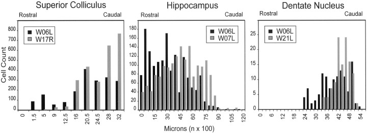
Fig. 3.
Distribution of second-order labeled neurons. The histograms show the rostrocaudal distribution of labeling in the superior colliculus for W06L (Multi) versus W17R (LIP) (left), in the hippocampus for W06L (Multi) versus W07L (7a) (middle), and in the dentate nucleus for W06L (Multi) versus W21L (7b) (right).

