
Fig. 2.
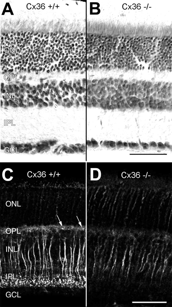
Retinal structure and immunoreactivity for connexin36 in wild-type (Cx36 +/+) and Cx36-deficient (Cx36 −/−) retinas. Vertical sections of Cx36 +/+ (A) and Cx36 −/− (B) retina counterstained with toluidine blue show no obvious differences in their overall structure. When immunolabeled with antibodies to Cx36, paraformaldehyde-fixed, frozen vertical sections of wild-type retina display the typical strong, punctate staining pattern in the inner plexiform layer and a weaker, punctate staining pattern (arrows) in the outer plexiform layer (C) that cannot be detected in the retinas of Cx36 −/− animals (D). Nonspecific staining of transretinal fibers is present in both Cx36 −/− and Cx36 +/+ mice and is attributable to paraformaldehyde fixation. ONL, Outer nuclear layer; OPL, outer plexiform layer; INL, inner nuclear layer;IPL, inner plexiform layer; GCL, ganglion cell layer. Scale bars, 50 μm.