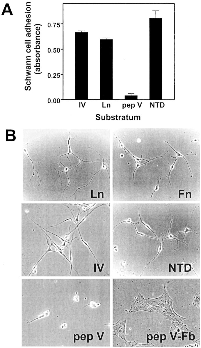
Fig. 4.

Schwann cell adhesion and spreading on ECM proteins. A, Schwann cells were plated on dishes coated with the indicated ECM proteins and incubated in serum-free medium with heregulin. Schwann cell adhesion was measured after 3 hr using the crystal violet assay. Values shown are mean ± SD for three separate measurements. B, Schwann cells or fetal rat fibroblasts were plated on dishes coated with the indicated ECM proteins and incubated in serum-free medium with heregulin. Schwann cell spreading was monitored by phase contrast microscopy.Ln, Laminin; Fn, fibronectin;IV, collagen type IV; pep V, pepsinized collagen type V; pep V-Fb, rat embryo fibroblasts plated on pepsinized type V collagen.
