Fig. 8.

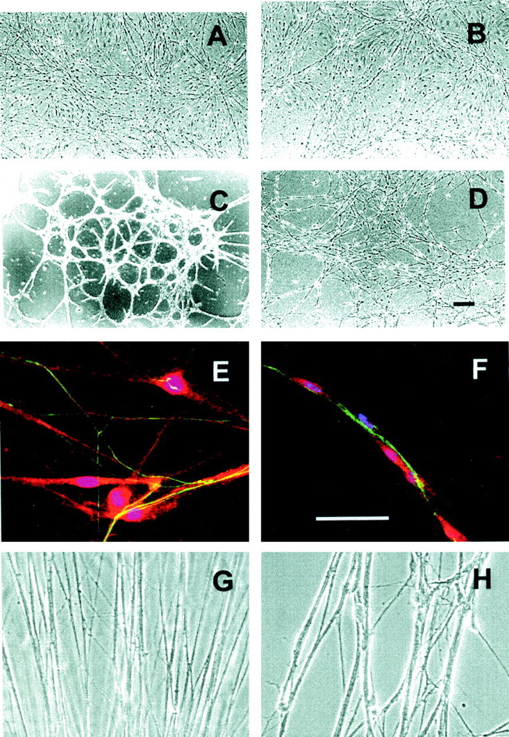
Organization of Schwann cell–axon units on ECM-coated surfaces. A–F, Dissociated rat embryo dorsal root ganglion neurons and Schwann cells were plated on dishes coated with collagen type IV (A, E), collagen type I (B), collagen type VSC (C, F), or uncoated plastic (D). The cells were cultured in serum-free medium with nerve growth factor for 4 d. A–D, Low-power phase contrast images.E, F, Higher-power views of cultures stained with anti-neurofilament antibodies (green, stains axons), anti-S100 antibodies (red, stains Schwann cells), and 4′,6-diamidino-2-phenylindole (blue, stains Schwann cell nuclei). (Note that superimposition of blueand red produce magenta.) Scale bars, 100 μm. G, H, Dorsal root ganglion explants were placed on dishes coated with laminin (G) or laminin plus collagen type VSC (H) and cultured in serum-free medium with nerve growth factor for 4 d. Axons were photographed using phase contrast optics near the site where they emerged from the ganglia.
