
Fig. 1.
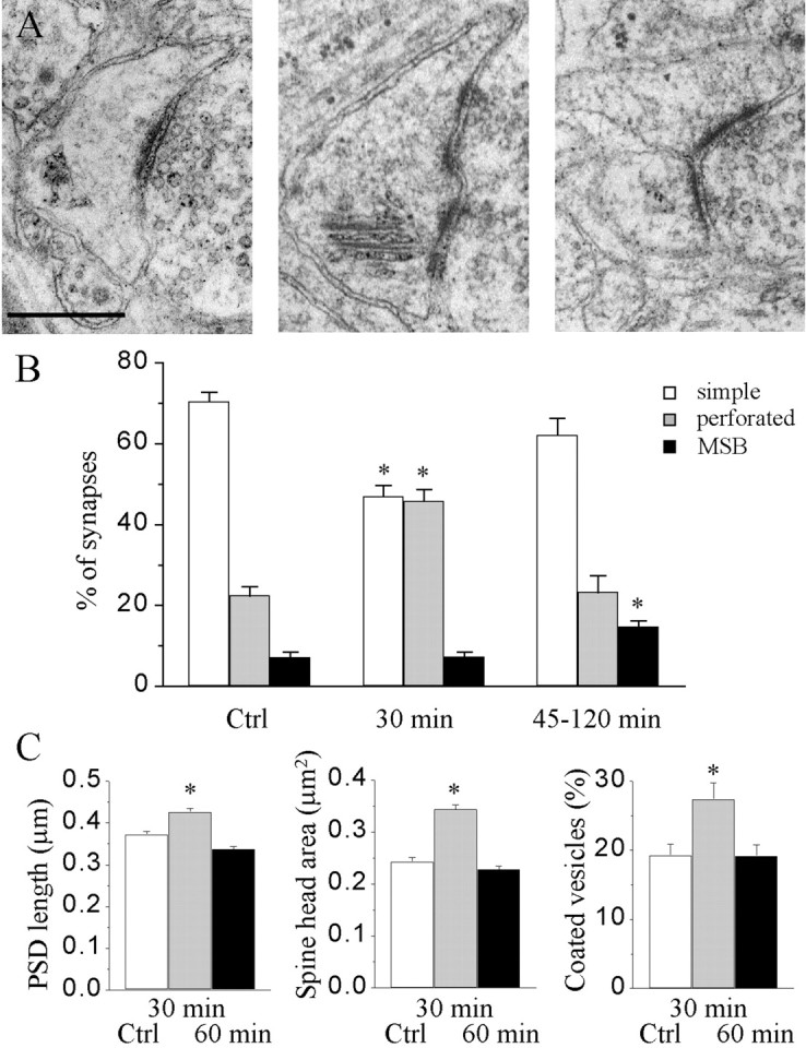
Changes in the proportion of different types of synapses and their characteristics during LTP. A, Illustration of a simple synapse with a single PSD (left), a synapse with a perforated PSD (middle), and an MSB (right). Scale bar, 0.5 μm. B, Proportion of the three types of synapses under control conditions and at 30 and 45–120 min after LTP induction (n = 4–14 hippocampal slice cultures and 358–1519 synaptic profiles analyzed; *p < 0.01).C, Changes in PSD length (left), spine head profile area (middle), and the proportion of spine profiles containing coated vesicles (right) determined via single-section analysis of the entire population of labeled synapses (n = 4–9 hippocampal slice cultures and 360–660 synaptic profiles; *p < 0.01).Ctrl, Control.