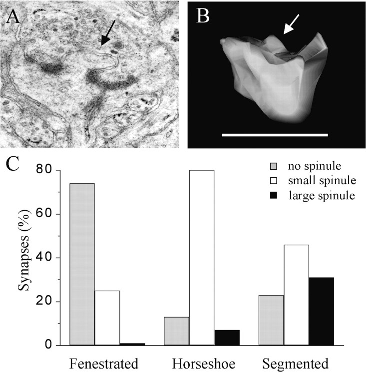
Fig. 5.
Presence of large spinules associated with the PSD of synapses with segmented PSDs. A, Illustration of a spine profile with a large spinule emerging between the two parts of the PSD. B, Illustration of a spinule on another reconstructed spine; arrows in A andB point to large spinules. Scale bar, 1 μm.C, Proportion of synapses with fenestrated, horseshoe-type, and segmented PSDs that exhibited no spinules (gray column) or spinules of small (<0.2 μm;white column) or large (>0.2 μm; black column) size in a population of 15, 15, and 13 reconstructed synapses, respectively.

