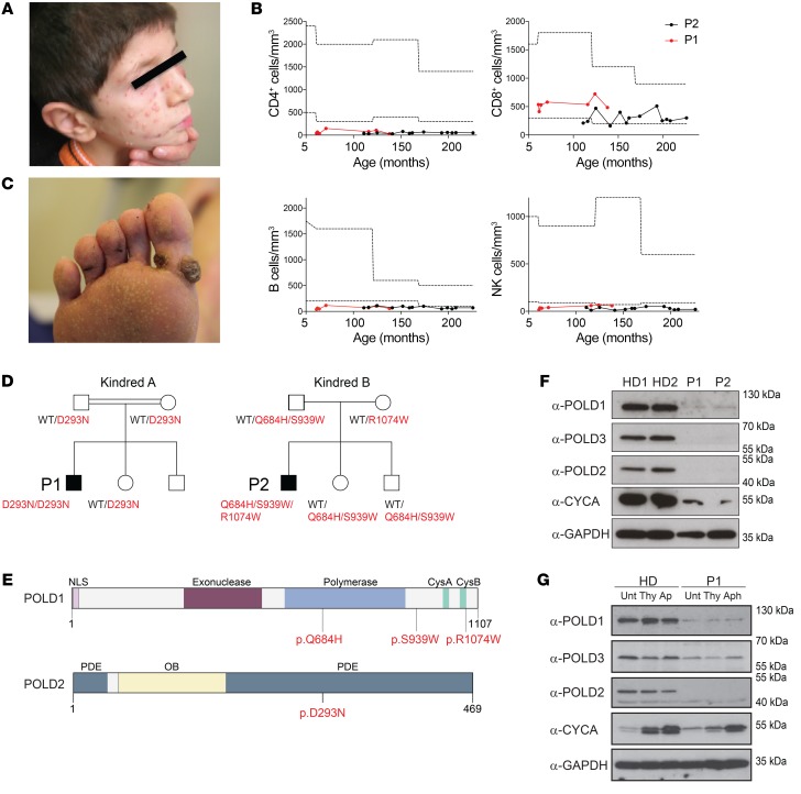
Figure 1. Identification of hypomorphic mutations affecting the polymerase δ complex in patients presenting with syndromic combined immunodeficiency.
(A) Molluscum contagiosum skin infection in P1. (B) Longitudinal peripheral blood CD4+ T cell, CD8+ T cell, B cell, and NK cell counts in P1 and P2. Dotted lines represent the range of reference values. (C) Viral skin warts in P2. (D) Familial segregation of the identified POLD1 and POLD2 mutations in the families of P1 and P2, indicating an autosomal-recessive pattern of inheritance. (E) Domain structure of POLD1 showing the polymerase domain, the exonuclease domain, the nuclear localization signal (NLS) domain, and the cysteine-rich, metal-binding domains CysA and CysB. Domain structure of POLD2 depicting the PDE and oligonucleotide binding (OB) domains. Mutation sites are indicated in red. (F) Protein levels of POLD1, POLD2, POLD3, and CYCA in PBMCs after anti-CD3 and anti-CD28 stimulation for 48 hours. See complete unedited blots in the supplemental material.(G) Protein levels of POLD1, POLD2, and POLD3 in primary fibroblasts. GAPDH was used as a loading control. Cells were untreated (Unt) or synchronized by double-thymidine (Thy) treatment or aphidicolin (Aph) treatment for 24 hours. α, anti.

