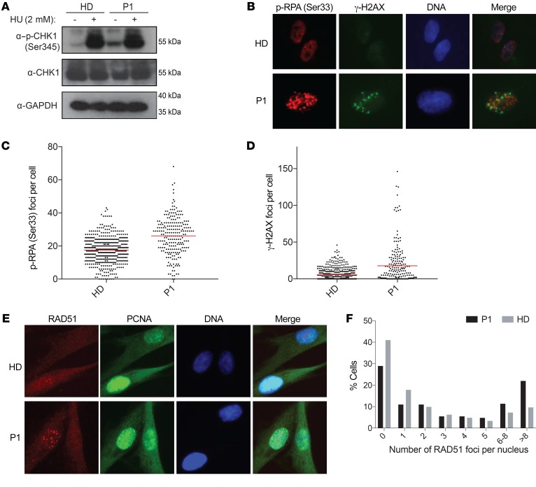
Figure 4. Polymerase δ mutation leads to replication-related DNA lesions associated with activation of the S-phase checkpoint.
(A) Immunoblot analysis of CHK1 phosphorylation at Ser345 in HD and P1 fibroblasts upon treatment with 2 mM hydroxyurea (HU). Total CHK1 and p-CHK1 were run on 2 different gels. GAPDH was used as a sample-processing control. (B) Immunofluorescence staining of p-RPA (Ser33) and γH2AX in HD and P1 fibroblasts. Original magnification, ×40. (C) Quantification of p-RPA (Ser33) foci per nucleus. Average of p-RPA (Ser33) foci: 17.48 (HD) and 26.11 (P1). (D) Quantification of γH2AX foci per nucleus. Average of γH2AX foci: 5.61 (HD) and 17.71 (P1). Number of cells counted in C and D: 481 (HD) and 223 (P1). (E) Immunofluorescence analysis of RAD51 foci and PCNA (S-phase marker) in HD and P1 fibroblasts. Original magnification, ×40. (F) Quantification of RAD51 foci per nucleus. Number of cells counted: 415 (HD) and 273 (P1). Image analysis was performed using CellProfiler, version 2.0.

