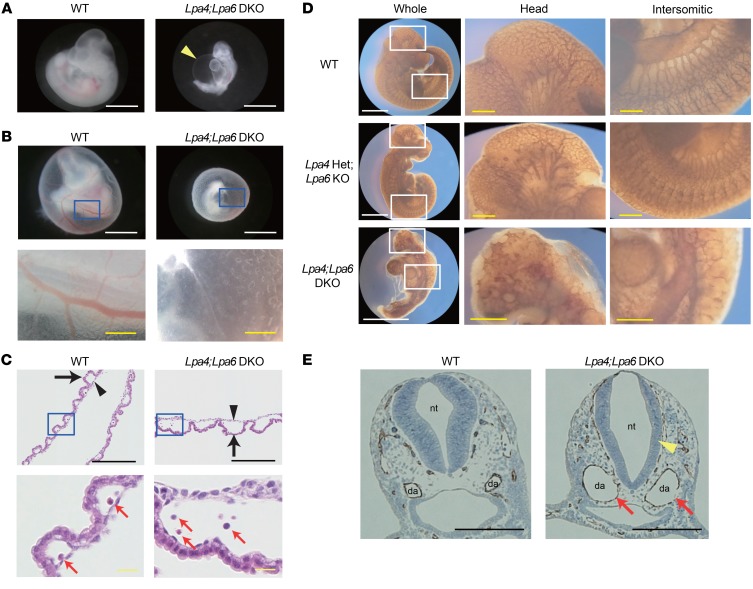
Figure 1. Lpa4;Lpa6-DKO embryo proper and yolk sac have vascular abnormalities.
(A) Gross morphology of embryo proper at E10.5. Lpa4;Lpa6-DKO embryo proper had pericardial effusion as indicated by an arrowhead (WT, n = 11; DKO, n = 22 ). Scale bars: 1 mm. (B) Gross morphology of yolk sac at E10.5. The bottom panels show higher-magnification images of the boxed areas indicated in the top panels. Lpa4;Lpa6-DKO yolk sac lacked large and branched blood vessels (WT, n = 9; DKO, n = 21). White scale bars: 1 mm; yellow scale bars: 200 μm. (C) H&E-stained transverse section of yolk sac. The bottom panels show higher-magnification images of the boxed areas indicated in the top panels. Endoderm (arrow) and mesoderm (arrowhead) of Lpa4;Lpa6-DKO yolk sac were more widely separated (WT, n = 5; DKO, n = 8). Erythrocytes (red arrows) were present in DKO yolk sacs. Black scale bars: 1 mm; yellow scale bars: 200 μm. (D) Vascular networks of embryo proper at E10.5. Blood vessels were stained with anti–PECAM-1 antibody. The middle and right panels show higher-magnification images of the boxed areas indicated in the left panels. Poor vascular networks in the head and intersomitic regions of Lpa4;Lpa6-DKO embryo proper were noted (WT, n = 4; Lpa4-Het;Lpa6-KO, n = 4; Lpa4;Lpa6-DKO, n = 5). White scale bars: 1 mm; yellow scale bars: 200 μm. (E) Transverse sections of the jugular area of embryo proper at E9.5. Blood vessels were stained with anti–PECAM-1 antibody. Enlargement of dorsal aortae (red arrows) and thinning of neural tube wall (yellow arrowhead) were remarkable in Lpa4;Lpa6-DKO embryo proper (WT, n = 5; DKO, n = 6). da, dorsal aorta; nt, neural tube. Scale bars: 200 μm. Representative images are shown.

