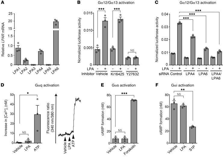
Figure 3. LPA4 and LPA6 in HUVECs mediate LPA-induced Gα12/Gα13 activation.
(A) Expression of mRNA for LPA receptors was detected by RT-qPCR. (B) SRF-RE-Luc reporter assay was performed to detect Gα12/Gα13–Rho activation. LPA (10 μM, 6 hours) increased the reporter activity, which was attenuated by the ROCK inhibitor Y27632 (10 μM, 10 minutes pretreatment) but not by the LPA1/LPA3 antagonist Ki16425 (10 μM, 10 minutes pretreatment). (C) LPA-induced SRF-RE-Luc activity was attenuated by LPA4/LPA6 siRNAs (12.5 nM each, 48 hours pretreatment). (D) Intracellular calcium influx assay was performed to detect Gαq activation. A representative trace of changes in intracellular Ca2+ concentration ([Ca2+]i) is shown on the right. HUVECs were unresponsive to LPA (10 μM). Adenosine 5′-triphosphate (ATP; 10 μM) was used as a positive control, because ATP evokes calcium influx via P2Y receptors that predominantly couple with Gαq. (E) cAMP assay was performed to detect Gαs activation. No significant increase in cAMP level was observed in HUVECs treated with LPA (10 μM). Forskolin (20 μM) was used as a positive control. (F) cAMP assay was performed to detect Gαi activation. Inhibitory effect of LPA (10 μM) on cAMP accumulation stimulated by forskolin (20 μM) was not observed in HUVECs. Sphingosine-1-phosphate (S1P; 100 nM) was used as a positive control. Data are mean ± SEM of triplicates. *P < 0.05, **P < 0.01, ***P < 0.001, 1-way ANOVA followed by Tukey’s multiple-comparisons test.

