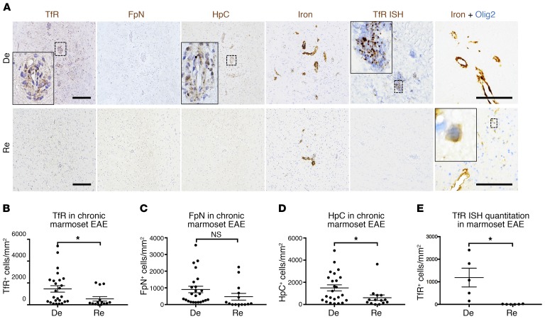
Figure 5. Remyelinated marmoset EAE lesions have lower levels of iron-regulating proteins than demyelinated lesions.
(A) Immunohistochemical staining for representative demyelinated (De) and remyelinated (Re) chronic (>6 weeks old) EAE lesions show higher levels of transferrin receptor (TfR) and hepcidin (HpC) in demyelinated compared with remyelinated lesions. However, both remyelinated and demyelinated lesions harbor iron. In remyelinated lesions, iron can be found inside Olig2+ oligodendrocyte-lineage cells. Quantification of (B) TfR, (C) ferroportin (FpN), (D) HpC, and (E) TfR mRNA shows that demyelinated lesions have higher numbers of TfR+ and HpC+ cells per unit area, as well as higher TfR mRNA expression. Dots represent individual lesions. *P < 0.05 (2-tailed t test). Scale bars: 100 μm. Counterstain: hematoxylin for single-stained slides. Lesions selected from marmosets 1, 2, and 6.

