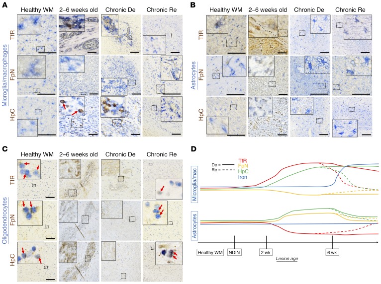
Figure 6. Iron-regulating protein changes are cell-specific and dynamic in healthy white matter and lesions.
(A) Panel of double stains (Iba1 for microglia/macrophages, together with the iron-regulating proteins transferrin receptor (TfR), ferroportin (FpN), and hepcidin (HpC)) in healthy white matter and various stages of marmoset EAE lesions. Microglia/macrophages weakly express all 3 iron-regulating proteins in healthy white matter. As lesions age, TfR and HpC levels increase, remaining high in chronically demyelinated (De) lesions but returning to baseline in remyelinated (Re) lesions. On the other hand, FpN levels slightly drop during demyelination but also return to normal upon remyelination. (B) Panel of double stains (GFAP for astrocytes, together with the same iron-regulating proteins). In the healthy white matter, astrocytes express TfR, which is lost upon demyelination but returns with remyelination. FpN and HpC levels increase in 2- to 6-week-old and chronically demyelinated lesions but also return to baseline with remyelination. (C) Panel of double stains (Olig2 for oligodendrocyte-lineage cells, likely a mixture of oligodendrocyte precursor cells and mature oligodendrocytes, together with iron-regulating proteins). In healthy white matter, TfR, FpN, and HpC are all expressed in the oligodendrocyte lineage (red arrows). In 2- to 6-week-old and chronically demyelinated lesions, oligodendrocyte-lineage cells are not detected. In repaired/remyelinated lesions, repopulated oligodendrocyte-lineage cells show all 3 proteins at relatively normal levels (red arrows). (D) Summary of iron regulation changes in microglia/macrophages and astrocytes during marmoset EAE lesion development and repair. Scale bars: 100 μm. Lesions selected from marmosets 1, 2, 6, 8, and 10.

