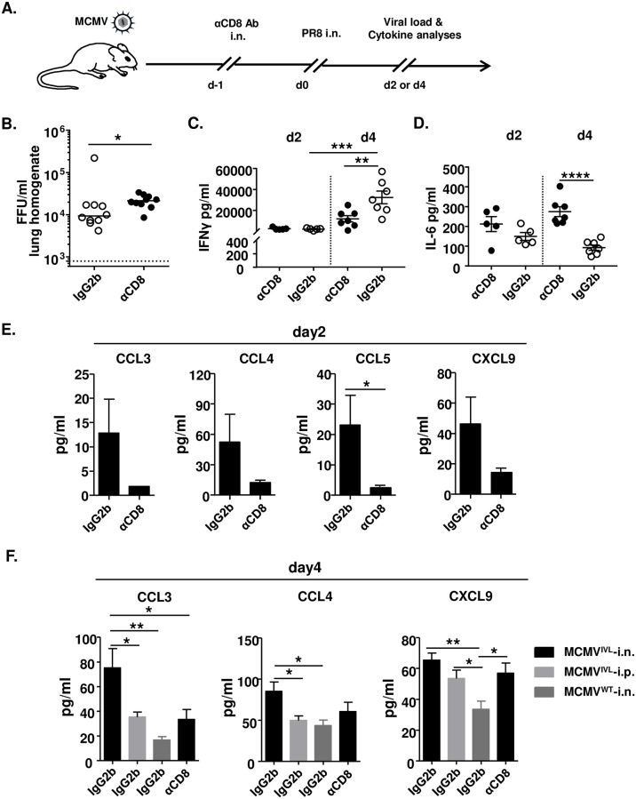
Fig 5. CD8TRM cells facilitate the elimination of IAV.
BALB/c mice were i.n. or i.p. immunized with 2 x 10⁵ PFU MCMVIVL or i.n. with MCMVWT. During latency (> 3 months p.i), mice were treated with αCD8 or IgG2b antibodies and challenged with IAV (PR8) (i.n., 1100 FFU). Leukocytes were isolated from lungs on day 4 post-challenge for flow cytometric analysis. (A) Graphic representation of the mucosal CD8+ T cell depletion protocol. (B) IAV titers in the lungs on day 4 post-challenge of MCMVIVL i.n. immunized mice. Two independent experiments were performed and pooled data are shown. Each symbol represents one mouse, n = 10. Group medians are shown. (C-F) The concentration of inflammatory cytokines and chemokines were measured in the BALF on day 2 and day 4 post-IAV challenge. (C) The concentrations of IFNγ and (D) IL-6 in the BALF of each MCMVIVL i.n. immunized mice are shown as symbols. Group means +/- SEM are shown. (E) The concentration of CCL3, CCL4, CCL5 and CXCL9 in the BALF on day 2 post-challenge. (F) The concentration of CCL3, CCL4 and CXCL9 in the BALF on day 4 post-challenge. Two independent experiments were performed and pooled data are shown. n = 5–7. Bars indicate means, error bars are SEM. Significance was assessed by Mann-Whitney U test, One-way ANOVA test, or Two-way ANOVA test. *P<0.05, **P<0.01, ***P<0.001, ****P<0.0001.

