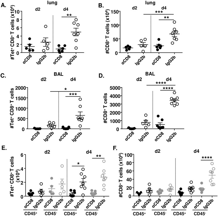
Fig 6. CD8TRM cells facilitate the accumulation of CD8+ T cells in the lungs.
BALB/c mice were immunized with 2 x 10⁵ PFU MCMVIVL via the i.n. route. During latency (> 3 months p.i), mice were i.n. treated with αCD8 (black circle;grey circle) or IgG2b (black-lined circle;grey-lined circle) antibodies and challenged with IAV (PR8) (i.n., 1100 FFU) one day after. Anti-CD45 antibodies were injected intravenously 3–5 min before euthanasia. Leukocytes were isolated from lung tissue to analyze the CD8+ T cell response on day 2 and day 4 post-challenge. (A) Total cell counts of IVL-specific CD8+ T cells of both intravitally labeled (CD45+) or unlabeled (CD45-) in the lung tissue. (B) Total cell counts of CD8+ T cells of both CD45+ and CD45- in the lung tissue. (C) Cell counts of IVL-specific CD8+ T cells in the BAL. (D) Cell counts of total CD8+ T cells in the BAL. (E) IVL-specific CD8+ T cells or (F) Total CD8+ T cells that were intravitally labeled or remained unlabeled in the lung tissue were counted on day 2 or day 4 post IAV challenge. Two independent experiments were performed and pooled data are shown. Each symbol represents one mouse, n = 5–7. Group means +/- SEM are shown. Significance was assessed by One-way ANOVA test. *P <0.05, **P <0.01, ***P <0.001, ****P <0.0001.

