. 2019 Sep 16;8:e46773. doi: 10.7554/eLife.46773

Figure 1. Nxph4 expression marks the components of select brain circuits.

(A–D) β-galactosidase staining of adult Nxph4βgeo/+ mice shows signals in mammillary body-related circuits (A), circumventricular organs (B–C), and cerebellar-vestibular circuits (D). Blue staining represents β-galactosidase activity. Top panels are stereotaxic maps adapted from the Paxinos and Franklin mouse brain atlas, with the red circle indicating the region for the image shown on the bottom panels. Aiv, Biv, Ciii, and Div illustrate the main connections among Nxph4+ regions in each circuit described. The inset in Di shows the blue staining in the cerebellar cortex granular layer. Scale bars: Ai, 500 μm; Aii, 100 μm; Aiii, 500 μm; Bi 500 μm; Bii, Biii 200 μm; Ci, Cii 100 μm; Di, Dii, Diii, 1 mm; Di inset, 100 μm. SUM, supramammillary body; MM, medial mammillary body; LM, lateral mammillary body; DTN, dorsal tegmental nucleus; PrS, presubiculum; LH, lateral hypothalamus; SFO, subfornical organ; MPO, median preoptic nucleus; CNX, nucleus of vagus nerve; AP, area postrema; DCN, deep cerebellar nuclei; MV, medial vestibular nucleus; SV, superior vestibular nucleus; ECu, external cuneate nucleus; Psol, parasolitary nucleus. (E) Double in situ staining of adult wild type mouse DCN and cerebellar cortex with probes against Nxph4, Vglut2 (an excitatory neuron marker), and Gad1 (an inhibitory neuron marker). PC: Purkinje cells. Scale bars: 50 μm. (F) Double in situ staining of mouse cerebellum shows that Nxph4 signals overlap with Grm2 (a Golgi cell marker) but not Pvalb or Calb2. Scale bars: 200 μm.

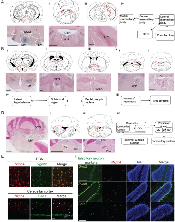
Figure 1—source data 1. Brain regions expressing Nxph4.
DOI: 10.7554/eLife.46773.004

Figure 1.

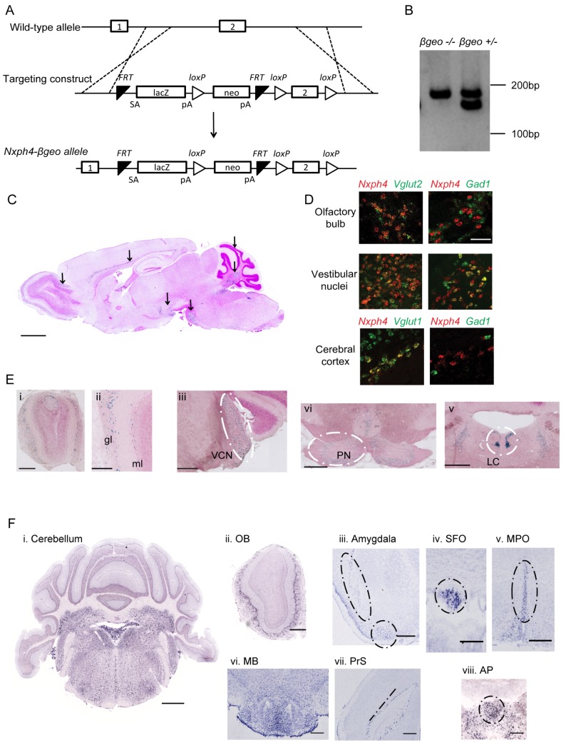
Figure 1—figure supplement 1. Generation of Nxph4-βgeo knock-in mouse and characterization of Nxph4 expression by β-galactosidase staining and in situ hybridization.

Figure 1—figure supplement 1.

(A) Targeting scheme to generate Nxph4-βgeo allele. Doted lines indicate homologous recombination sites. ES cells with correct Nxph4-βgeo allele were obtained from KOMP. (B) Genotype analysis of tail DNA showed a 180 bp band from Nxph4 wild type allele and a 150 bp band from Nxph4-βgeo allele. PCR products were separated on 3% MetaPhor agarose gel. (C) A parasagittal section of an adult Nxph4βgeo/+ mouse brain processed for β-galactosidase staining, resulting in a blue reaction product where Nxph4 is expressed. Arrows indicate regions with strong Nxph4 expression. Scale bar: 1 mm. (D) Double in situ staining of adult wild type mice with probes against Nxph4, Vglut1/2 (excitatory neuron markers), and Gad1 (an inhibitory neuron marker). Nxph4 co-localized with Vglut2 in the olfactory bulb, vestibular nuclei, and cerebral cortex, but also showed overlapping signal with Gad1 in the vestibular nuclei. Scale bar, 100 μm. (E) β-galactosidase staining of adult Nxph4βgeo/+ mice shows Nxph4 expression in the glomerular layer (gl) and mitral cell layer (ml) of the main olfactory bulb, ventral cochlear nucleus (VCN), pontine nuclei (PN), and locus coeruleus (LC). Scale bars: Eii, 200 μm; others, 500 μm. (F) RNA in situ hybridization with a probe against Nxph4 detected Nxph4 in several regions of adult mouse brain. Scale bars: Cerebellum, 1 mm; OB (Olfactory Bulb), 500 μm; Amygdala, 100 μm; SFO, 200 μm; MPO, 200 μm; MB, 300 μm; PrS, 100 μm; AP, 100 μm.