. 2019 Sep 16;8:e46773. doi: 10.7554/eLife.46773

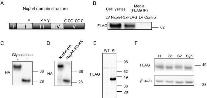
Figure 3. Nxph4 is a glycosylated protein that can be detected in the synaptosomes.

(A) A domain model of Nxph4 (adapted from Missler and Südhof, 1998). I: signal peptide; II: a variable domain; III: a conserved domain; IV: a linker region; V: C-terminal domain. Positions of N-glycosylation sequences are marked by letter Y, and the conserved cysteine residues are identified by the letter C. (B) Immunoblotting of samples from cultured cortical neurons that are infected with lentivirus expressing Nxph4-3xFLAG or the control lentivirus. Nxph4-3xFLAG was detected in the cell lysates as well as the media. (C) Treatment with glycosidase altered the electrophoretic motility of recombinant Nxph4-HA. (D) Nxph4-HA-4Q mutant has a smaller molecular mass compared with the wild type recombinant Nxph4-HA. (E) Immunoblotting analysis detects Nxph4-3xFLAG expression in the KI mouse synaptosomes. (F) Immunoblotting analysis of fractions derived from cerebellar synaptosomal preparation detects Nxph4-3xFLAG in the synaptosomes. β-actin was used as loading control. H: homogenate. S1 and S2 are successive supernatants in the synaptosomal preparation protocol. S2 is also the cytosolic fraction. Syn: synaptosomes.

Figure 3.

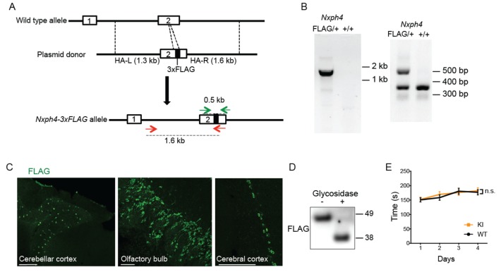
Figure 3—figure supplement 1. Generation and characterization of Nxph4-3xFLAG knock-in mice.

Figure 3—figure supplement 1.

(A) A schematic diagram of the strategy to generate an Nxph4-3xFLAG knock-in allele. An sgRNA is designed to excise the coding region corresponding to the C-terminal region of Nxph4 third domain. In the donor vector, the 3xFLAG sequence is inserted after the sequence of Nxph4 third domain. The homologous arms of the donor vector are indicated as HA-L and HA-R. Two pairs of primers designed for genotyping are shown with green and red arrows. (B) Representative genotyping results using two different pairs of primers to distinguish Nxph4FLAG/+ from Nxph4+/+ mice. Left, the forward primer is outside of the HA-L and the reverse primer in within 3xFLAG region (red arrows in A). Right: the forward and the reverse primers flank the 3xFLAG sequence (green arrows in A). (C) Representative immunofluorescence images illustrate the expression of Nxph4-3xFLAG in specific brain regions. Scale bars: 200 μm. (D) Treatment with glycosidase altered the electrophoretic motility of Nxph4-3xFLAG. (E) Latency to fall from the accelerating rod (mixed male and female mice, n = 17–19). Data are presented as mean ± SEM. n.s., not significant; by two-way ANOVA.
Figure 3—figure supplement 2. Validation of synaptosomes preparation.
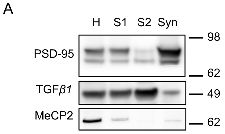
Figure 3—figure supplement 2.

(A) Immunoblot analysis of fractions derived from different steps of synaptosomal preparation. Synapse specific protein PSD-95 showed enriched signal in purified synaptosomes as expected. Somatic protein TGF-β1 is enriched in the cytosolic fraction S2. Nuclear protein MeCP2 is barely detectable in S1, S2 or Syn. H: homogenate. S1 and S2 are successive supernatants in the synaptosomal preparation protocol. S2 is also the cytosolic fraction. Syn: synaptosomes.