. 2019 Sep 16;8:e46773. doi: 10.7554/eLife.46773

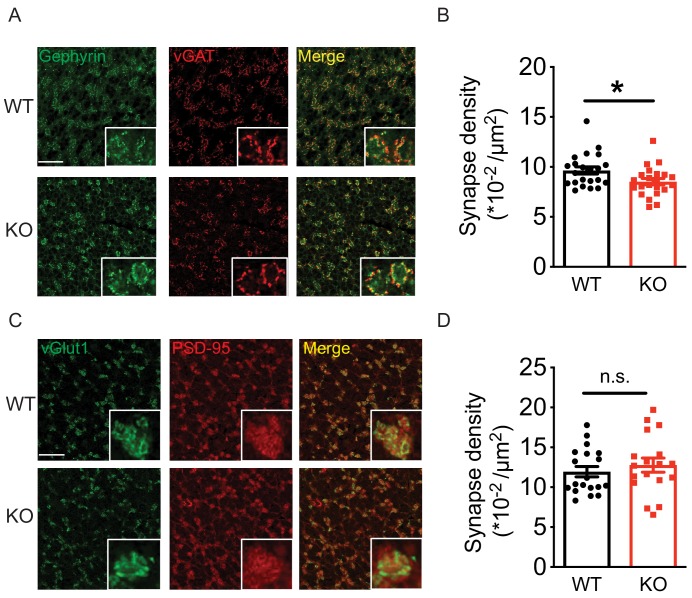
Figure 7. Loss of Nxph4 reduced Golgi-granule inhibitory synapse number.

(A) Gephyrin and vGAT staining in the cerebellar granular layer indicates Golgi-granule inhibitory synapses. (B) Quantification of puncta co-expressing gephyrin and vGAT as an indicator of inhibitory synapse number. (C) vGlut1 and PSD-95 staining in the cerebellar granular layer indicates mossy fiber-granule cell excitatory synapses. (D) Quantification of puncta co-expressing vGlut1 and PSD-95 as an indicator of excitatory synapse number. Data are presented as mean ± SEM. n.s., not significant; *p<0.05; by t test (B) or Mann-Whitney U test (D).

Figure 7.

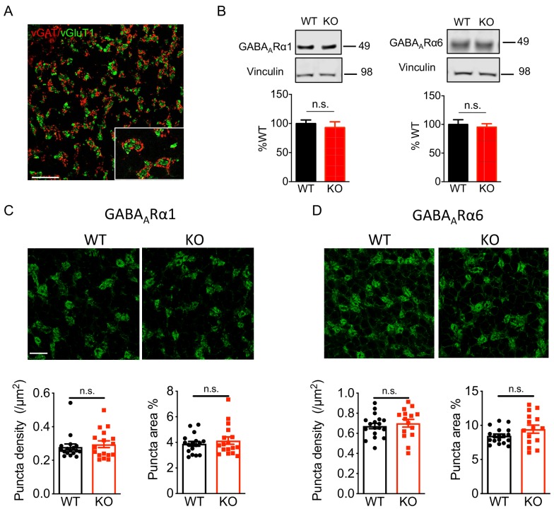
Figure 7—figure supplement 1. Nxph4 KO mice showed normal expression and localization of GABAARs.

Figure 7—figure supplement 1.

(A) In the cerebellar granular layer, vGAT marks the Golgi cell inhibitory terminals and vGlut1 marks the mossy fiber endings. Scale bar: 20 μm. (B) Immunoblotting analysis of GABAARα1/6 in the cerebellar synaptosomes of the WT and Nxph4 KO mice (n = 6–8). (C–D) Top: Immunofluorescence staining of GABAARα1 (C) and GABAARα6 (D) in the cerebellar granular layer. Scale bar: 20 μm. Bottom: Quantification of GABAAR cluster density and size (n = 17 sections; 3 mice). Data are presented as mean ± SEM. n.s., not significant; by t test.