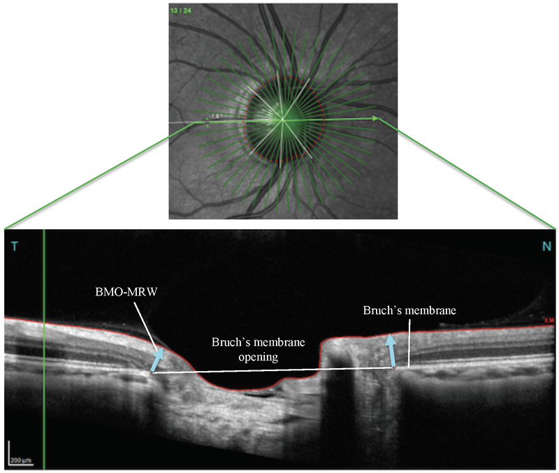
Fig. 1.
Panel A depicts a Spectralis optical coherence tomography (OCT) derived fundus image showing the optic disc with corresponding retinal microvasculature, as well as 24 radial slices of the peri-papillary region and 3 circular scans at 3.5 mm, 4.1 mm, and 4.7 mm diameters surrounding the optic disc (Panel A). The corresponding OCT image (Panel B) shows the Bruch’s membrane opening – minimum rim width (BMO-MRW) flanking the optic cup (blue arrows). Scans were acquired from a healthy participant

