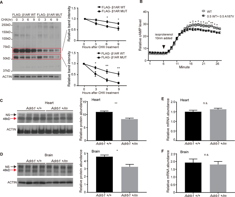
Figure 2. The β1AR A187V mutation alters protein stability and cAMP production.
(A) Degradation assay of β1AR in transfected HEK293 cells. Twenty-four hours after transfection, cells were treated with 100 μg/ml CHX and harvested at indicated time points. Bands inside the two red boxes indicate β1AR protein of different sizes in the SDS gel. Quantified results are shown on the right. β1AR protein levels at the starting point (t=0 hours) were normalized to 1.
(B) The β1AR A187V mutation confers altered downstream signaling output in response to isoproterenol in cultured cells. Heterozygous expression of β1AR A187V and WT leads to a reduction in cAMP production.
(C and D) Western blotting results of endogenous β1AR protein from the heart (C) and brain (D) lysates of Adrb1+/+, and Adrb1+/m animals. N=4 mice per group. NS, non-specific band. Quantified results are shown on the right.
(E and F) q-RTPCR results of Adrb1 mRNA normalized to Actin mRNA from the heart (E) and brain (F) tissues of Adrb1+/+ and +/m animals. N=4 mice per group.
* P<0.05, **P<0.01, n.s.= not significant. Two way RM ANOVA, post-hoc Sidak’s multiple comparisons test for (A) and (B). Two-tailed Student’s t-test for (C)-(F). Error bars represent ± SEM.

