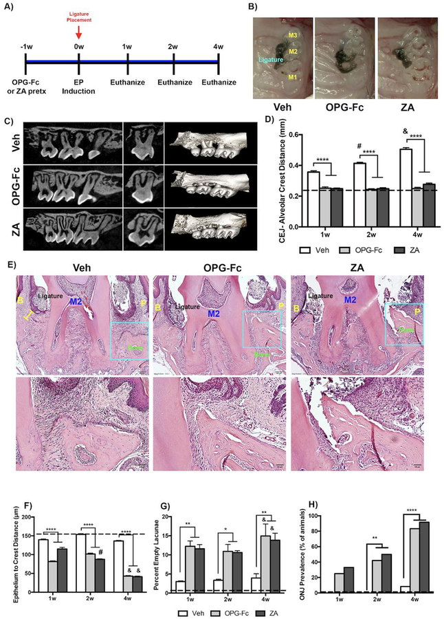
Figure 1: Radiographic and Histologic Measurement of Veh, OPG-Fc, and ZA treated animals.
A) Experimental Timeline. B) Clinical photomicrographs of Veh, OPG-Fc or ZA treated animals. C) Parasagittal and coronal cross sections and 3-dimensional μCT reconstructions of Veh, OPGFc or ZA animals at the 4-week time point D) Quantification of the CEJ-Alveolar Crest Distance for Veh, OPG-Fc, and ZA treated animals, at the 1-week, 2-week, and 4-week time points. E) Representative coronal H&E images of Veh, OPG-Fc, and ZA treated animals at the 4 weeks after ligature placement. F) Quantification of the epithelium to crest distance. G) Quantification of the percent empty osteocytic lacunae. H) ONJ Prevalence at the 4-week time point. The yellow bar denotes the Epithelium to Alveolar Crest Distance. Data represents mean value ± SEM. **** = statistical significance p<0.0001, *** = statistical significance p<0.001, ** = statistical significance p<0.01, * = statistical significance p<0.05. % statistical significance p<0.05 vs 1w. & = statistical significance p<0.05 vs 1w and 2w. # = statistical significant vs. 1w. M1= First Molar, M2= Second Molar, M3= Third Molar. B= Buccal; P=Palatal.

