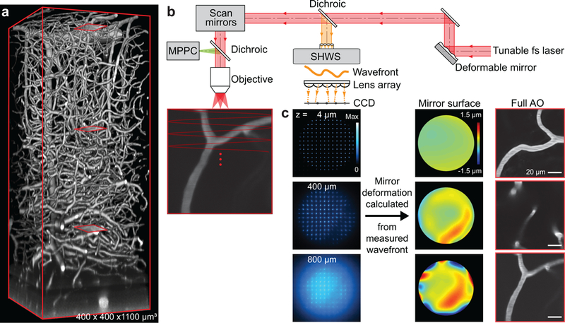
Figure 1. Aberration measurements up to 800 μm below the pia in parietal cortex of awake mouse via wavefront sensing from a microvascular-based guide star.

(a) In vivo two-photon imaging of vasculature in a 400×400×1100 μm3 volume of cortex with the blood plasma labeled with Cy5.5 conjugated to 2MD-dextran. The excitation wavelength was λ = 1.25 μm. Red boxes show three example sub-regions used for wavefront sensing in panel c.
(b) Adaptive optical two-photon microscopy based on direct wavefront sensing from the descanned signal of the guide star, formed by two-photon excitation of Cy5.5 in the vasculature lumen, with a Shack-Hartmann wavefront sensor. The wavefront is corrected by a deformable mirror. The signal for brain imaging is detected by the multi-pixel photon counters.
(c) Spot pattern (left) formed by the wavefront sensor from the descanned guide star signal; the position of each spot away from its center determined the tilt of the wavefront. Wavefront maps (center) reconstructed from the wavefront sensor spots patterns at different depths. Microvessels (right) at 14, 400, and 800 μm below the pia (red boxes in panel a) form the guide stars by two-photon excitation.
