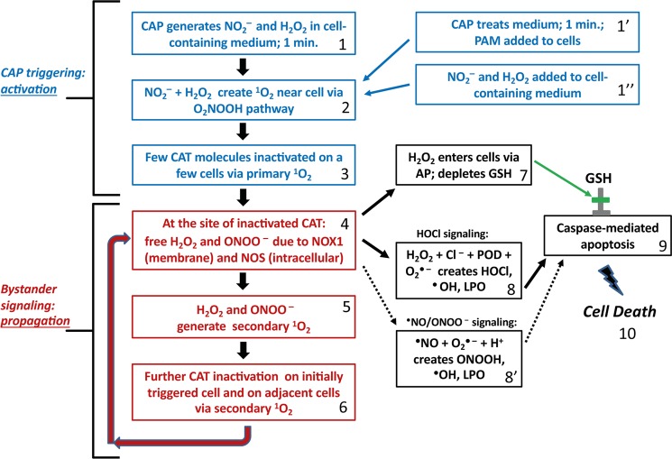
Figure 2.
Flow chart of major steps in CAP leading to selective apoptosis of tumor cells. Step 1: CAP generates NO2− and H2O2 in cell containing medium for 1 minute. Alternatively, CAP is used to treat medium, creating PAM (step 1′). Defined concentrations of NO2− and H2O2 containing medium are used in reconstitution experiments (step 1”). Step 2: NO2−- and H2O2 create primary 1O2 near cells following O2NOOH pathway, as described in Figure 1. Step 3: Few catalase molecules on a few cells are inactivated due to primary 1O2 near cells. Step 4: At the site of inactivated catalase, H2O2 and ONOO− (generated through NOX1 and NOS) are no longer decomposed. Step 5: The reaction between H2O2 and ONOO− is leading ultimately to secondary 1O2. Step 6: This additional 1O2 leads to further catalase inactivation and the process cycles back to step 4. Step 7: Increased H2O2 resulting from catalase loss from secondary 1O2 leads to H2O2 entering cells via aquaporins, leading to antioxidant glutathione depletion. Step 8: In parallel with step 7, increased H2O2 resulting from catalase loss from secondary 1O2 also leads to HOCl generation by peroxidase, in the presence of Cl−. The interaction between NOX1-derived O2●− leads to ●OH formation near the cell membrane and lipid oxidation. Step 8′: If HOCl signaling is suppressed, an alternative ●NO/ONOO− signaling can also lead to lipid peroxdiation. Step 9: If both lipid peroxidation and glutathione depletion occurs, then caspase-associated apoptosis can take place, finally leading to cell death. Steps 1–3 correspond to CAP triggering or activation of a few cells, thereby initiating propagating bystander signaling in steps 4–6. Steps 7–9 are the steps that lead to the final cell apoptosis. These steps are activated only if the repeated performance of steps 4–6 has caused a sufficiently high degree of catalase inactivation for reactivation of HOCl or ●NO/ONOO− - mediated apoptosis-inducing signaling.

