Figure 1.
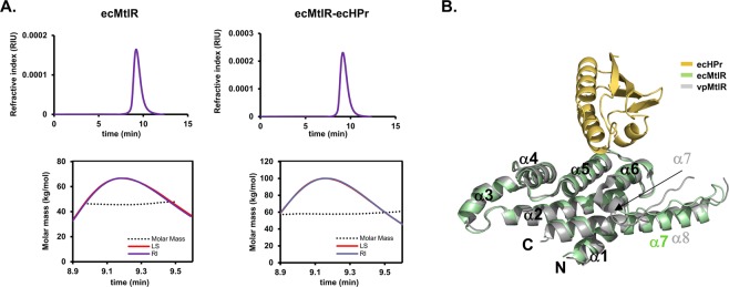
E. coli MtlR and HPr forms a stable heterotetramer. (A) The SEC-MALS analysis of ecHisMtlR (left) and the ecHisMtlR-HPr complex (right). Refractive index (RI) and light scattering (LS) signals were measured to determine protein concentration (top panels) and molar mass (bottom panels), respectively. (B) Structure of the ecMtlR monomer (green) in complex with HPr (yellow). The vpMtlR structure11 (PDB 3BRJ, gray) is superposed on the ecMtlR structure. N- and C-termini and α-helices of both MtlRs are labeled. vpMtlR has an additional α-helix (α7 in gray) compared with ecMtlR, thus α8 of vpMtlR in gray and α7 of ecMtlR in green are structurally aligned.

