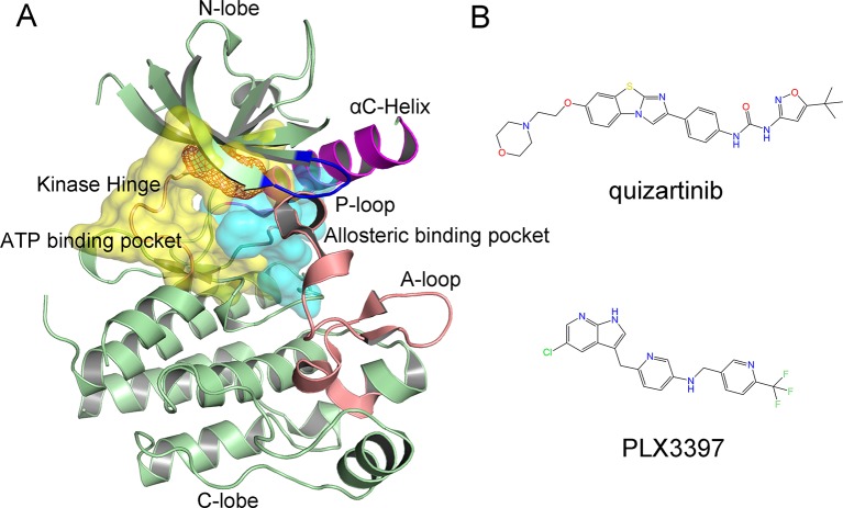
Figure 1.
Crystal structure of FLT3 (PDB code: 4RT7) and representative inhibitors. (A) Overview of the Mps1 structure, F691L gatekeeper mutation is colored orange dots. The ATP binding pocket is colored yellow surface and allosteric binding pocket is colored cyan surface; (B) 2D chemical structures of quizartinib and PLX3397.

