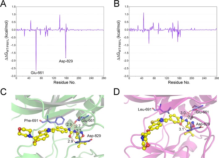
Figure 6.
The energetic differences of the residue contributions to the binding free energies between the WT system and F691L system (∆∆G=∆G WT − ∆G F691L). (A) FLT3-WT + quizartinib and FLT3-F691L + quizartinib; (B) FLT3-WT + PLX3397 and FLT3-F691L + PLX3397; (C) representative structure of FLT3-WT + quizartinib; (D) representative structure of FLT3-F691L + quizartinib. Hydrogen bonds are colored red, and the key residues are colored purple.

