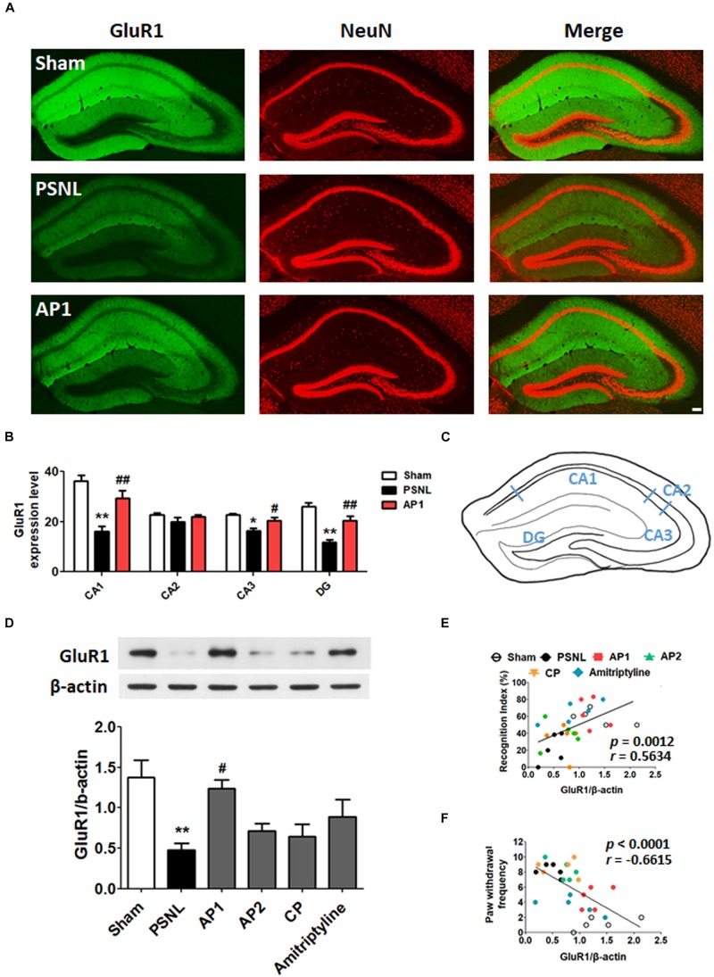
FIGURE 6.
Effects of acupuncture on the expression levels of hippocampal GluR1 receptor. These results show the changes in hippocampal GluR1 expression levels (CA1, CA2, CA3, and DG) after administration of acupuncture (AP1, AP2, or CP) or amitriptyline (10 mg/kg, i.p.) for 28 consecutive days (A–F). Free-floating coronal hippocampus sections from the three groups (Sham, PSNL, and AP1) were subjected to immunofluorescence with GluR1 (green) and NeuN (red) antibodies to label GluR1-positive NeuN in neurons (A). Representative graphs showing the expression levels of GluR1 in the hippocampus (A,B) and a representative figure showing the hippocampal regions in the mouse brain (C). n = 3–4/group. Scale bar: 100 μm. ∗p < 0.05, ∗∗p < 0.01 compared to the Sham group in each area. #p < 0.05, ##p < 0.01 compared to the PSNL group in each area. The expression levels of hippocampal GluR1 protein were examined (D). n = 5/group. ∗∗p < 0.01 compared to the Sham group. #p < 0.05 compared to the PSNL group. All data were analyzed with a one-way ANOVA followed by Newman–Keuls post hoc tests. All data are expressed as the mean ± SEM. The cognitive function values in the NOR test and nociceptive values in the von Frey test were correlated with the GluR1 protein levels in the hippocampus (E,F). The r-values were analyzed with the Spearman rank correlation coefficient. AP1, acupuncture 1 (GB30 and GB34); AP2, acupuncture 2 (HT7 and GV20); CP, control point.

