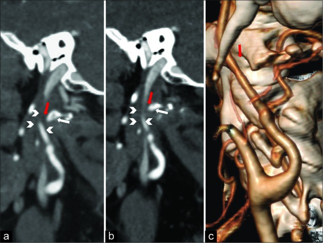
Figure 2:

Postoperative computed tomography angiography examination. (a and b) Sagittal multiplanar reconstruction demonstrating the surgically generated distance (red arrows) between the reshaped C1 transverse process (white arrows) and internal carotid artery (ICA) (arrowheads). (c) Volume rendering technique reconstruction image showing the new anatomical relationship between ICA and C1 transverse process (red arrows).
