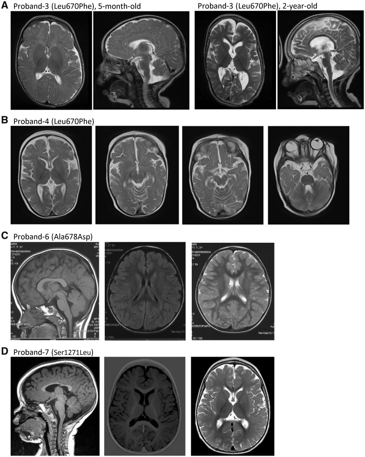
Figure 2.
Brain MRI of patients with GRIN2D variants. (A) Proband 3 (Leu670Phe) showed cortical atrophy, global loss of white matter volume and enlargement of the lateral ventricles; compare MRI at 5 months old (left two panels) to 2 years old (right two panels). (B) Proband 4 (Leu670Phe) showed mild cortical atrophy. (C) Proband 6 (Ala678Asp) had normal MRI left: sagittal T1. middle: coronal T2. right: coronal T2 at 3 years 5 months of age. (D) Proband 7 (Ser1271Leu) had normal MRI left: sagittal T1, middle: coronal T1, right: coronal T2 at 14 months old.

