
Fig. 3.
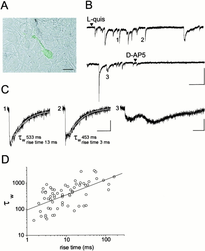
Stimulation of astrocytes results in glutamate-mediated, fast activation of NMDARs in the sensor cell.A, NR-GFP cell plated on cultured astrocytes. Scale bar, 10 μm. B, TheNR-GFP cell shown in A responded with repetitive, inward currents after stimulation of astrocytes with 10 μm quisqualate. Holding potential, −55 mV. Calibration: 20 pA, 10 sec. C, Events 1,2, and 3 are reported at expanded scales. Calibration: 5 pA, 500 msec. Rise time value of the current and decay time constant τw of the curve best describing the decay are reported. The fitting curve (white line) is superimposed to the currents. D, Scatter plot of rise time values of the currents recorded from 13 NR-GFPcells expressed as a function of the current decay time constant τw (mean ± SEM; rise time, 20.2 ± 3.75 msec; range, 1.5–167 msec; τw 563 ± 74.9 msec; range, 37–2896 msec; amplitude, 20.2 ± 6.7 pA; range, 3.2–272 pA;n = 65). Very slow currents (rise and decay times >200 msec and 3 sec, respectively) such as event 3, were not included in our sample. The line is the linear fit to the data (r = 0.61; p < 0.0001).